Dans la même rubrique
-
Partager cette page
Device for shearing tissue [Offre de technologie]
The technology in a nutshell
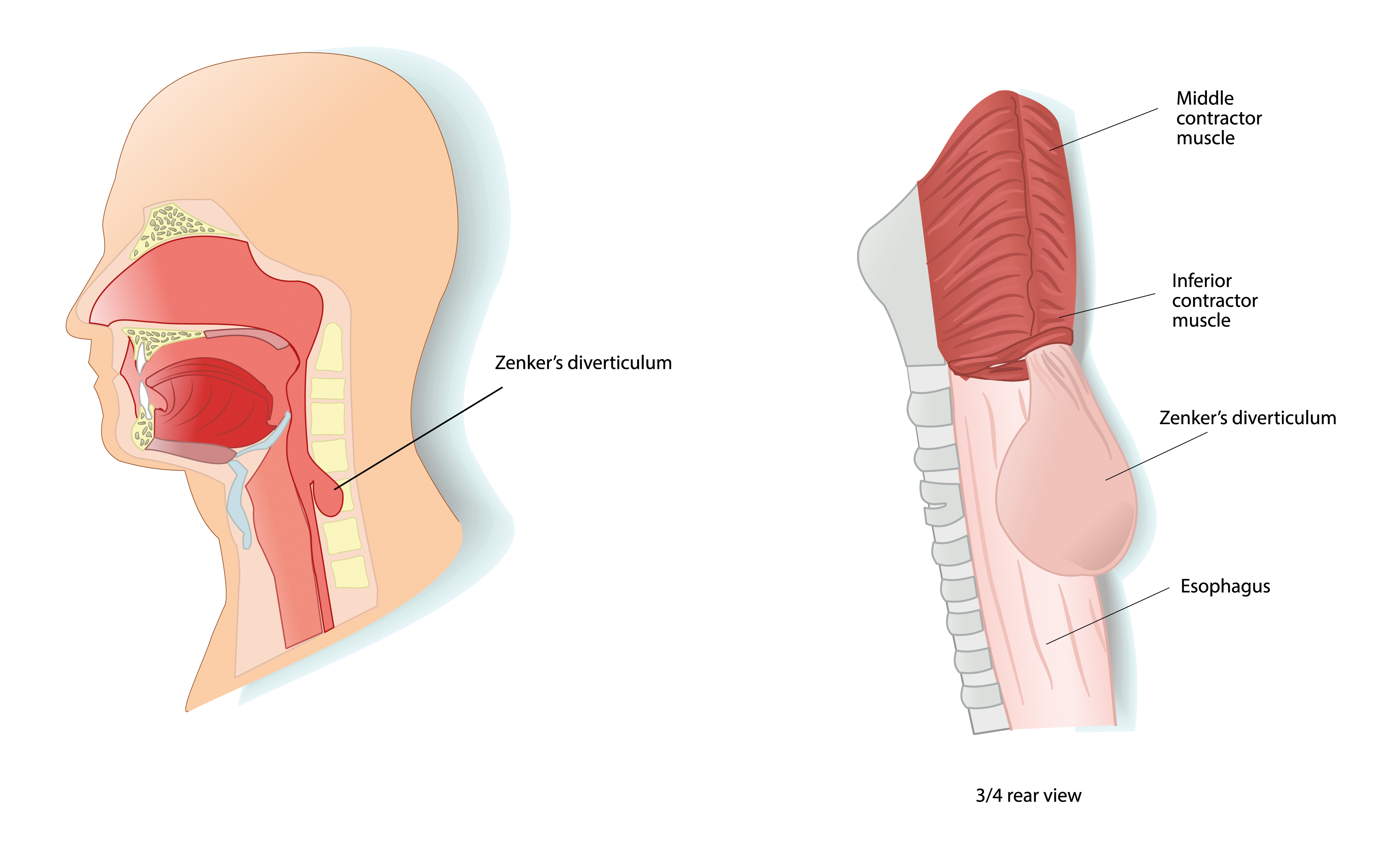
New magnets used as endoscopic devices for surgical procedures (mainly for people suffering from non-Zenker’s esophageal diverticulum).
State of the art
There exist a number of medical conditions in which a wall of an internal body cavity pouches out to form an undesired hollow protrusion, referred to as a diverticulum. Diverticula can appear in various body cavities, including but not limited to the gastrointestinal tract (esophagus, intestine, colon), the bladder, and the heart.
In some cases (e.g. non-Zenker’s esophageal diverticulum), the diverticulum can become the main pathway for food, inducing dysphagia and regurgitation and leading to significant weight loss and poor quality of life as the patient is no longer able to eat solid food. Current treatment techniques involve surgery with high rates of mortality (5%) and morbidity (20%). Endoscopic resection is also an option in some cases of diverticulum, but it is still scarcely used and highly complicated. Lastly, in other medical conditions, there is a need to open or adapt a septum between two cavities.
It is therefore desirable to provide a method and a device that enable creating an opening between adjacent cavities with an easy, atraumatic and endoscopic method.

The invention
The invention consists in an implant composed of magnets linked together by a wire. Through natural ducts—such as the esophagus—and using endoscopic tools, the magnets are positioned over the tissue to be cut (such as the wall between the esophagus and a diverticulum). The magnets strongly compress the tissue thus trapped, cutting it off from its blood supply. This results in tissue necrosis between the magnets and scarification at the edges, preventing an open wound. At the same time, the wire linking the magnets exerts a compression force on the remaining tissue, due to gravity affecting the magnets or to any other system creating a tension, resulting in a slow and atraumatic cutting of the tissue.


Key advantages of the technology
- The core of the invention is the wire linking the magnets, which serves as a cutting tool. Once the tissue trapped between the magnets has necrotized, the wire gently cuts the remaining wall by shearing tissue due to the magnets’ weight or another traction system.
- The use of a wire to act as cutting tool is the novel aspect. The invention’s main advantages are its non-invasiveness, a slow process in comparison with using a scalpel, and the variability in the size and shape of the cut.
Commercial interest
- Gastroenterology
- Laparoscopy
- Endoscopy
Technology Readiness Level
The inventors
Pr Alain Delchambre is Professor in the Biomed Group of the Bio-, Electro- And Mechanical Systems (BEAMS) department of the Faculty of Applied Sciences of the ULB.
The laboratories
For many years, there is a strong collaboration between the Faculty of Medicine, especially the Department of Gastroenterology, and the Bio-, Electro- And Mechanical Systems (BEAMS) department of the Faculty of Applied Sciences (School of Engineering) of the Université Libre de Bruxelles. This partnership has led to many research projects, patent applications and prototypes in the field of biomedical engineering.
The main interest of the Biomed group (BEAMS) is to develop and analyze medical devices in collaboration with clinicians. The research axes of the biomed group are: flexible mechanics, bioelectronics, biomechanics. This research group has developed close relationships with many medical departments in Europe.
Keywords
- Magnets
- Compression Anastomosis
- Wire
- Tissue necrosis
- Tension force
- Septum
Collaboration type
- Licence agreement
- Research cooperation
IP status
Publication number: WO2018158395 (A1)
Patent pending in US, Europe, Canada, Brazil, Japan
The inventors
- Dr Jacques Devière
- Pr Alain Delchambre
Contact
ULB Research Department
Joachim Ruol
Business developer
+32 (0)2 650 30 67
joachim.ruol@ulb.be

