In the same section
-
Share this page
Neurological Health Assessment by Peripheral Measurement of Brain Sensorimotor Oscillations [Technology Offer]
The technology in a nutshell
Method to record β-sensorimotor cortical oscillations based on wearable mechanical vibration sensors for assessment and monitoring of neurological health enabling to bypass the use of more expensive and complex EEG or MEG modalities.
State of the art
Sensorimotor cortex β-oscillations are the hallmark of sensorimotor brain functions, reflecting motor skills and neurological health. Their characterization usually requires electroencephalography (EEG) or magnetoencephalography (MEG) measurements with specialized and sometimes cumbersome equipment. Diagnosis and monitoring of Parkinson’s disease, monitoring of rehabilitation and recovery after cerebrovascular accident, assessment of severity of schizophrenia and monitoring response to intervention for autism spectrum disorders are all situations where easy, rapid and inexpensive measurement of β-oscillations would be of particular interest for the patients especially in regions with difficult access to EEG and/or MEG.
The invention
The present invention is a method and corresponding device to easily and rapidly measure β-band vibrations. It relies only on a vibration sensing unit comprising a force transducer possibly combined with a linear accelerometer. Owing to the simplicity of the setup, the method enables a swift and efficient monitoring of β-sensorimotor cortical oscillations. β-band vibrations signal may simply be recorded while the patient squeezes the vibration sensing unit or is performing a simple task such as changing the contraction force or performing an additional movement.
The technology is perfectly suited for remote diagnosis, monitoring and rehabilitation assessment of patients with motor brain dysfunctions or lesions.
Key advantages of the technology
- Fast, easy and reliable measure of brainwave beta rhythms not requiring EEG or MEG measurements
- Method based on simple, readily available wearable mechanical sensors
- Method perfectly suited for remote diagnosis and monitoring via telemedicine
Potential applications
- Diagnosis and monitoring of Parkinson’s disease
- Monitoring of rehabilitation and recovery after cerebrovascular accident
- Assessment of severity of schizophrenia
- Monitoring response to intervention for autism spectrum disorder
Technology Readiness Level

TRL-3 Proof of concept of the method in several applications.
The team
The Translational Neuro-Anatomy and Neuro-Imaging Laboratory (LN2T) is part of the ULB Neuroscience Institute (UNI) of Université libre de Bruxelles (ULB). The main fields of research are language, sensorimotor processes and learning/memory. This multidisciplinary laboratory translates neuroimaging research performed in healthy individuals into methods allowing to investigate the pathophysiology of brain disorders and to contribute to patients' clinical management.
The Laboratory has developed methods to investigate human brain function relying on different imaging modalities of its state of-the-art Neuroimaging platform including magnetoencephalography (MEG), high-density electroencephalography and PET-MR instruments and its strong expertise in neuroimaging data acquisition and signal processing.
The inventor
Mathieu BOURGUIGNON is Professor at the Université libre de Bruxelles (ULB) within the LN2T and the Laboratory of Neurophysiology and Movement Biomechanics. Physics engineer by training, his work is devoted to improving the understanding of brain mechanisms underlying motor control and speech processing, striving to develop applications in health and disease. His main research tools to probe sensorimotor control are MEG and EEG recordings, and human motion capture.

Figure
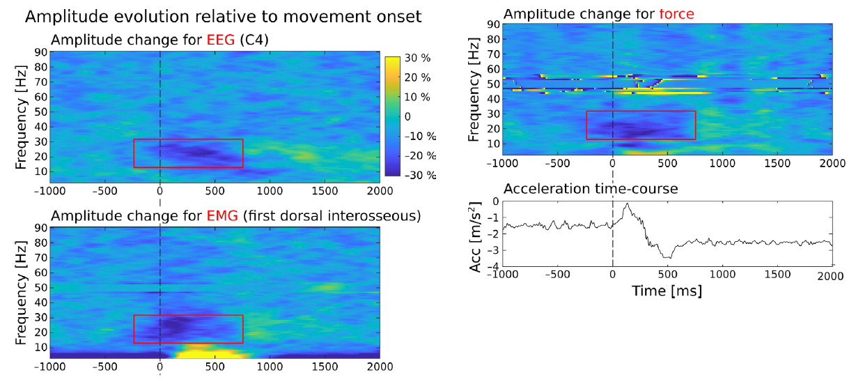
Time-frequency amplitude evolution of EEG (first Panel), EMG (second Panel) and measurements made using a vibration sensing unit (third Panel) in a task where intermittent elbow movements were performed while holding a steady pinch contraction. It is evident that, following movement onset (time 0), the sensorimotor cortical rhythm is attenuated, and that such attenuation is also visible in peripheral recordings. The last graph shows the corresponding acceleration time-course upon performing a movement.
Relevant publications
- MEG Insight into the Spectral Dynamics Underlying Steady Isometric Muscle Contraction, Journal of Neuroscience 2017, 37 (43), 10421
- Proprioceptive response strength in the primary sensorimotor cortex is invariant to the range of finger movement, NeuroImage 2023, 269, 119937.
Keywords
- β-Band cortical rhythms
- Wearable sensors
- Neurological health diagnosis and monitoring
Collaboration type
- Licence agreement
- R&D&I collaboration
IP status
- EP23168860.7
Priority Date: 20/04/2023 - PCT/EP2024/060326
- WO2024218103A1
Main inventor
Mathieu BOURGUIGNON
Contact
Knowledge Transfer Office
ULB Research Department
Joachim Ruol
Business developer
+32 (0) 496 75 94 93
joachim.ruol@ulb.be
